
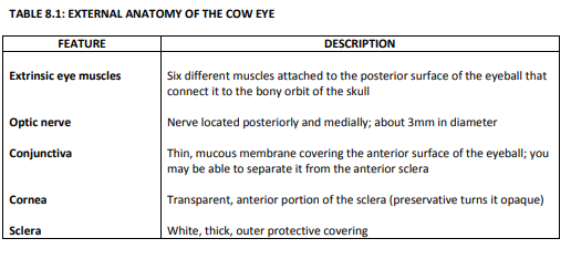
2.PNG
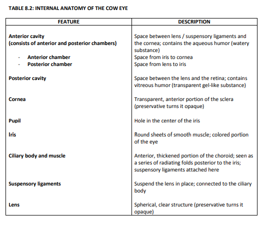
3.PNG
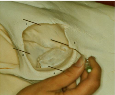
4.PNG
4 point block.PNG
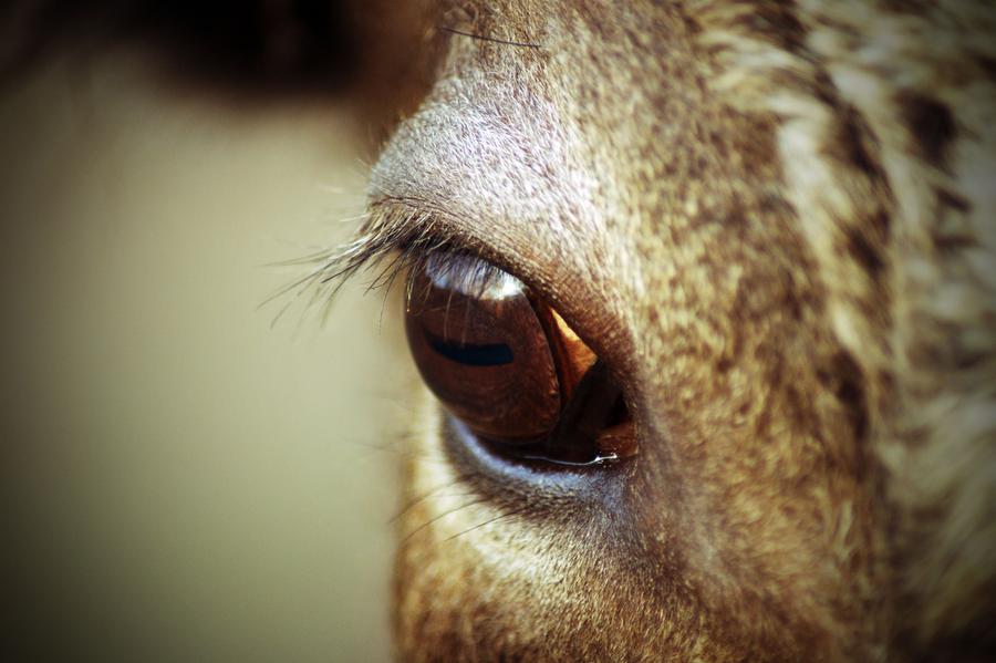
65e1fcab16faec60462623feef7dee02.jpg
Anatomy of the eye showing blood supply.png
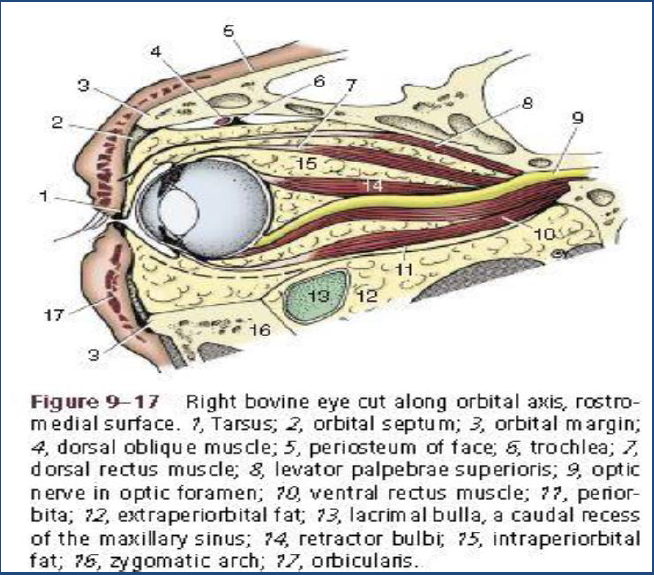
Anatomy of the periorbital structures.jpg
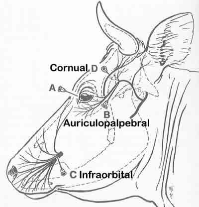
auriculopalpebral .jpg
Auriculopalpebral nerve block.url
Background 3EL.jpg
Background for anyone to use.jpg
background- laceration.jpg
Capture.PNG
Cherry Eye.jpg
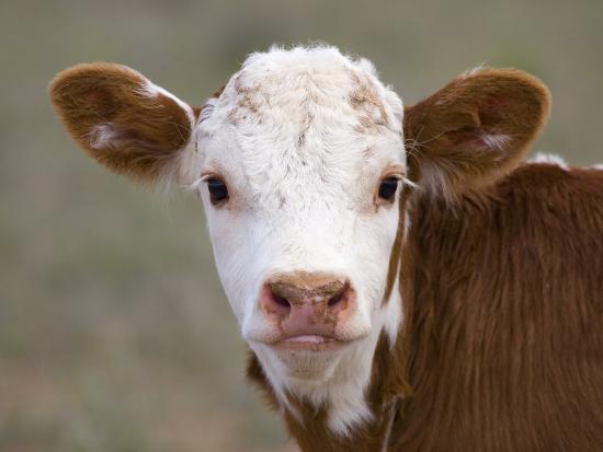
Cow.jpg
Cow 2.jpg
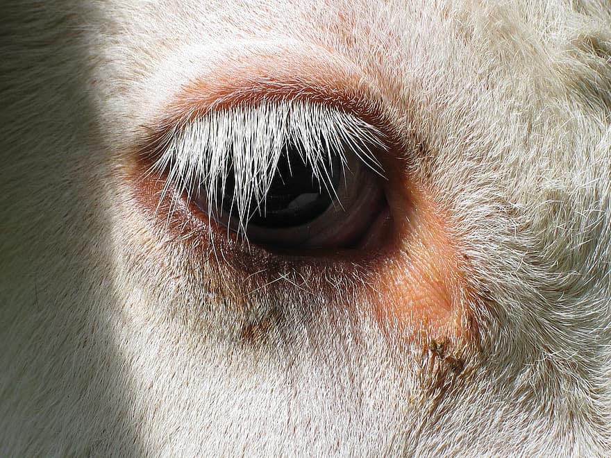
download.jpg
Drug and Calculations eye.pdf
Drugs and Calculations.pdf
Drugs and Calculations Eye .pdf
Exenteration Background.jpg
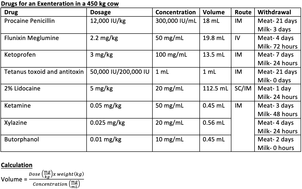
Exenteration Drug Table.png
EXENTERATION OF EYE BALL IN CATTLE - BOVINE OCULAR SQUAMOUS CELL CARCINOMA.url
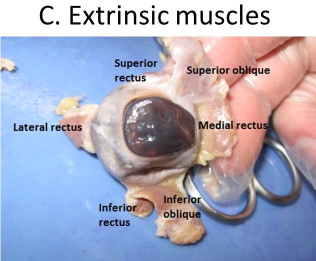
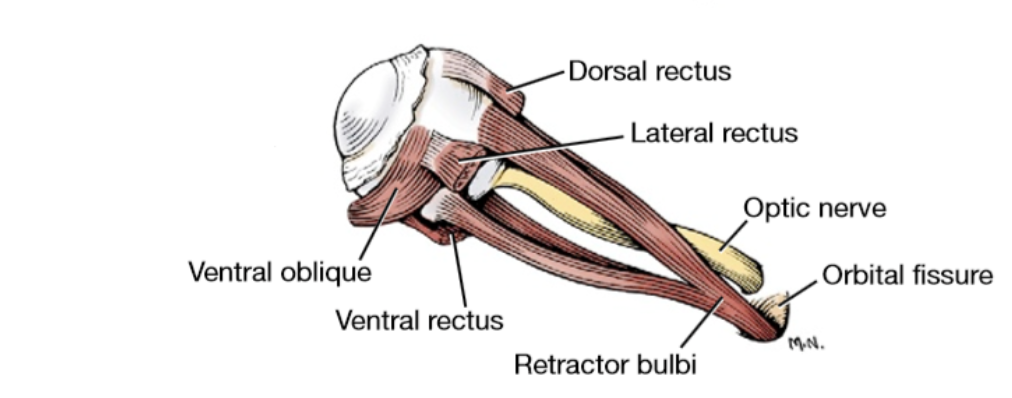
Extrinsic muscles of the eye.jpg
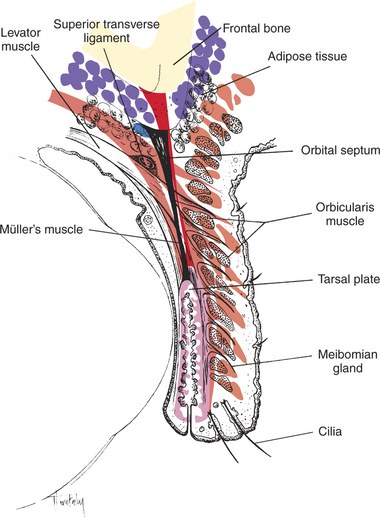
Eyelid anatomy.jpg
Eyelid Laceration Repair.url
Farmer Communication .pdf
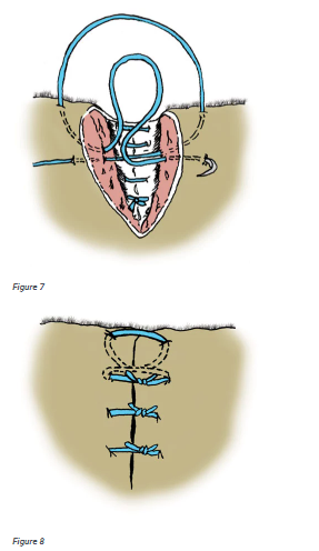
Figure 8 of suturing pattern.PNG
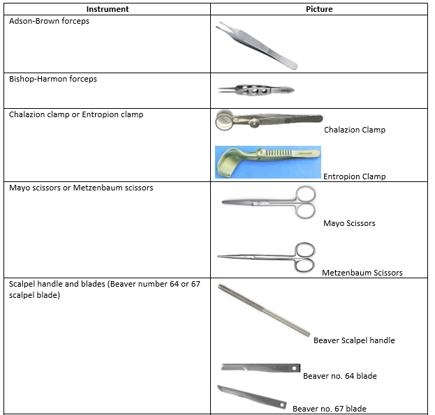
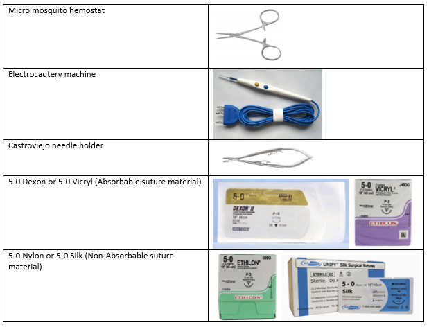
Instrument table 1.PNG
Instrument table 2.PNG
Lateral view of the muscles of the eye.png
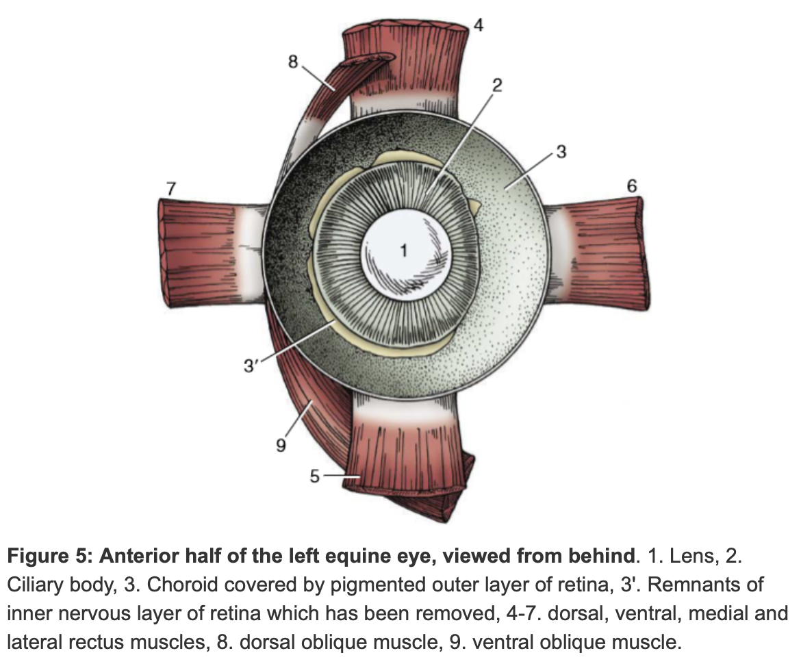
Muscles of the anterior half of the equine eye.png
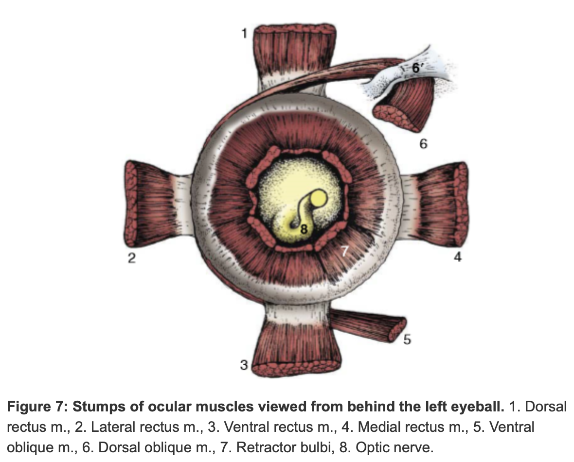
Muscles of the posterior half of the equine eye.png
Nerdy moo.jpeg
Nerve blocks image.png
Parts of the cow eye.png
Peterson's nerve block.url
Peterson-block-approach.jpg
Pre-op drugs.docx
restraint.jfif
retobulbar.url
Retrobulbar.url
SCC of the 3rd eyelid.jpg
Suturing of the muscle and subcutaneous tissue.PNG
Suturing of the skin.PNG
Third eyelid anatomy.jpg
Third eyelid removal.png
Third Eyelid Removal.url
Third eyelid removal 2.png